


Stretching: Understanding Flexibility, Mobility, and the Science Behind It


Stretching is considered essential for fitness and health, yet many misconceptions surround it. Flexibility and mobility are often confused – though one refers to passive range of motion, the other to active movement control. In today’s blog post, we take a detailed, science-based look at what stretching actually changes – and what it doesn’t.
Introduction
Stretching is one of the most universally accepted practices in health, fitness, and sport. From warm-ups and cool-downs to yoga sessions and physical therapy, it’s often seen as essential, a non-negotiable ritual for improving performance, preventing injury, and maintaining longevity.
But what if we’ve misunderstood what stretching really does?
Beneath the surface of daily routines lies a more complex truth – one grounded in biomechanics, neurophysiology, and material science. Because when we pull on muscles and feel that familiar tension, what’s actually changing? Is it the muscle itself? The joint? Our nervous system’s perception? Or nothing at all?
At the heart of this exploration is a common confusion between flexibility and mobility – terms used interchangeably but rooted in very different mechanisms. While flexibility is often treated as the end goal, mobility may be the real measure of movement. One is passive. The other is active, dynamic, and trainable.
What limits our range of motion isn’t always muscles. What feels like “lengthening” might just be a change in tolerance, not structure. And what we call progress might simply be temporary comfort, not long-term adaptation.
In the pages that follow, we’ll break down the mechanical and neurological layers of stretching. We’ll revisit terms like stiffness, extensibility, and viscoelasticity. We’ll look at how joints and tissues actually behave under load, and why most stretching protocols fall short of changing them. And most importantly, we’ll explore the evidence-based methods that truly improve movement.
This is not a quick-fix guide or a list of stretches. It’s a deeper investigation. A way to read between the lines of common practice and arrive at a clearer understanding of how our bodies respond to tension, perception, and time.
Let’s begin.
What is Stretching, Really?
At its core, stretching is the act of applying force to lengthen something. Be it a rubber band, a muscle, or connective tissue. In biomechanics, it’s essentially a tensile load: a pulling force that causes elongation in a structure. But while the act may seem simple, what’s happening inside the body is anything but.To understand stretching, it helps to borrow a lens from materials science. In a tensile test, a material is pulled from both ends to assess how it deforms. Engineers measure how much force is needed to create a given amount of strain (deformation), and how the material reacts. Whether it stretches elastically (returning to shape) or plastically (staying deformed). These principles apply remarkably well to biological tissues.


When we stretch a muscle or tendon, we’re essentially performing a biological tensile test. The tissue responds with deformation. But the amount and type of deformation depends on multiple variables: the tissue’s viscoelastic properties, hydration, temperature, rate of loading, and even its microscopic architecture.
In fitness and rehabilitation, stretching is usually aimed at increasing range of motion (ROM), often framed as improving “flexibility.” But here’s where things get murky: flexibility, as commonly used, is not a precisely defined physical property. It blends ideas from multiple disciplines – engineering, physiology, neurology – into a term that sounds intuitive but is scientifically vague.
From a material science standpoint, “flexibility” might mean a structure’s ability to bend or deform. In biomechanics, it more often refers to passive range of motion – how far a joint can move when external force is applied. But range of motion is not solely governed by the muscle’s mechanical limits. It’s also shaped by neurological factors like stretch tolerance, reflex activity, and pain perception. And just as importantly, it’s limited by the structural architecture of the body – the shape of bones, the orientation of joint surfaces, and the presence of surrounding connective tissues. For example, certain joints may reach a hard stop not because of muscles, but because of bony contact or joint design that restricts further movement.
So when someone says they’re “tight,” they may not be hitting a mechanical limit at all. They could be experiencing a protective neural response, a perception of threat, or even just unfamiliarity with a position. The tension isn’t just in the tissue, it’s in the nervous system’s response to it.
That’s why the classic assumption that stretching “lengthens” the muscle is often misleading. Most acute gains in flexibility result not from structural change but from a change in how the nervous system interprets stretch. It’s not that the muscle got longer, our body became more willing to let it go there.In short, stretching is a complex interaction between tissue mechanics and neural control. Understanding that sets the stage for rethinking how (and why) we stretch and what we should really be aiming to change.
The Language of Movement: Flexibility, Mobility, and Beyond
Stretching discussions often hinge on words that sound interchangeable but represent very different concepts. Chief among these are flexibility and mobility – terms that are often confused, even among professionals. But clarity here is crucial, because what we aim to change (and how we go about it) depends entirely on how we define these ideas.
Flexibility
In biomechanics, flexibility typically refers to the passive range of motion available at a joint – how far it can be moved by an external force without active muscular control. It’s influenced by the mechanical properties of tissues (like muscle and fascia), joint structure, and neural factors such as stretch tolerance.
In materials science, the term gets even trickier. Flexibility here can loosely describe how easily a material deforms under load. But more precise terms—like compliance (inverse of stiffness), ductility, and elasticity – are used to describe how a substance behaves when force is applied. These definitions don’t always transfer cleanly into human movement, which is why “flexibility” becomes a catch-all term that can obscure more than it explains.In casual use, “flexible” might mean bendable, adaptable, or limber but it doesn’t tell us why something bends or how it recovers afterward.
Mobility
Mobility is a more useful and precise term in the context of training and movement. It refers to a joint’s usable range of motion, especially under active control. This includes not just the capacity to get into a position, but the strength and coordination to control it.
Mobility can be:
- Active: The range of motion a person can access using their own muscular effort (e.g., lifting a leg without assistance).

- Passive: The range achieved when an external force moves the joint (e.g., a therapist stretching your leg).

While flexibility reflects the inherent characteristics of a tissue/material – its capacity to yield to an external force – mobility is what determines function. You may have flexible tissues that can easily deform under load, but without mobility, you can’t actively reach or control range. In other words, someone else (or some external force) might be able to push you into a certain position, but unless your nervous system and muscles can actively support and stabilize you in that position, it’s not a range you can truly use.
Other Overlapping Terms
- Extensibility: How far a tissue can stretch before it risks damage.
- Elasticity: The ability of tissue to return to its original shape after deformation.
- Stiffness: A material’s resistance to deformation. Often misunderstood as negative, but critical for force transfer and stability.
- Ductility: A material’s ability to deform plastically (i.e., not bounce back).
Understanding these distinctions helps us ask better questions:
Are we trying to change a tissue’s structure, or improve its control?
Are we limited by the nervous system, or by anatomical architecture?
Are we pursuing a range we can actually use, or just one that looks impressive in a static test?
Stretching only takes us so far. To move better, we must move more actively and more intelligently. That starts with speaking the right language.
Anatomy of a Limit: Joints and Muscles
When we talk about limits to range of motion, it’s tempting to blame tight muscles or stiff tissues. But human movement is governed not just by what can move, but by how the body is built to move. The architecture of the joints and surrounding tissues plays a crucial role in setting boundaries that stretching cannot override.
Joints: Structure Dictates Possibility
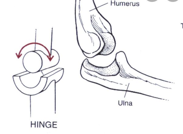
The structure of a joint including bone shape, orientation, and connective tissue arrangement sets the anatomical foundation for movement. This is the principle of form follows function. A ball-and-socket joint like the hip allows for a wide range of motion in multiple directions, while a hinge joint like the elbow is designed for flexion and extension within a much narrower arc.
Examples of Hip Joint and Elbow Joint:


Some joints are limited purely by soft tissues like ligaments, capsules, and muscle bulk. Others have bony restrictions that create a hard stop, regardless of how flexible the surrounding muscles might be. For example, elbow extension is limited by the olecranon process of the ulna contacting the humerus, while elbow flexion may be restricted by the approximation of muscle bellies.


These structural limits are non-negotiable. No amount of stretching will change the shape of your femur, deepen your hip socket, or alter the alignment of your vertebrae. Understanding joint anatomy helps us shift from trying to force range of motion to working within what’s realistically possible for a given body.
Muscles: Movers and Limiters
Skeletal muscles contribute both to mobility and stability. They generate force to move bones through space, but they also resist movement through stiffness and reflexive control. That stiffness is not just a passive resistance. It’s actively created by the muscle generating force, often in response to an external force. Reflexive control, such as the stretch reflex, triggers this force production automatically as a protective mechanism, increasing muscle activation to produce force and guard against unsafe movement.. A muscle’s resistance to stretch can come from two major sources:
- The contractile elements: the muscle fibers themselves.
- The non-contractile components: fascia, tendons, and connective tissue (such as ligaments and joint capsule)

Muscles don’t just limit motion through mechanical resistance. The nervous system plays a pivotal role in regulating how much range the body allows. One of the key players here is the stretch reflex: a fast, automatic response that resists sudden or excessive lengthening of a muscle (we’ll look at the muscle spindle later).
Muscle tone, tissue hydration, prior activity, and even emotional state can influence how “tight” or “stiff” a muscle feels. None of which are solved by stretching alone.
Connective Tissues: Springs with Memory
Tendons and ligaments behave like viscoelastic materials, they exhibit both elastic recoil and time-dependent flow. Their short-term response to stretching includes both immediate deformation and gradual adaptation, depending on load intensity, duration, and frequency. However, these adaptations are momentaneous and limited. The true game-changer for improvement isn’t passive stretching – it’s active loading that builds strength at end range.
In Summary
The body’s range of motion isn’t just about how stretchy your muscles feel. It’s the result of an integrated system: bones that form the framework, joints that dictate possible paths, muscles that generate and resist motion, and a nervous system that constantly calibrates safety and control. Stretching may nudge the limits temporarily, but long-term change requires training the system, not just tugging at it.
The Tissue Talks: Mechanical Properties of the Body
To truly understand what stretching does or doesn’t do, we need to listen to the tissues themselves. Muscles, tendons, ligaments, and fascia all respond to mechanical stress according to well-established principles of material science. These tissues don’t just stretch or resist; they deform, store energy, recoil, and adapt depending on their structural makeup and how they’re loaded.
Viscoelasticity: The Blend of Stretch and Flow
Most biological tissues are viscoelastic, meaning they exhibit both elastic and viscous behavior. Elastic materials (like a rubber band) return to their original shape after a load is removed. Viscous materials (like plasticine or honey) deform slowly and don’t return fully to their original shape. Viscoelastic tissues like muscle, tendon, and fascia do both; they stretch temporarily and also dissipate energy over time.


This explains why stretching a muscle feels different when done quickly vs. slowly. A fast stretch encounters more resistance (due to viscous response), while a slower stretch allows tissues to “creep” – time-dependent deformation under a persistent load – further as internal friction gives way. This property is central to why stretching needs time under tension to have any meaningful short-term effects and why it doesn’t create lasting change unless combined with loading.
Mechanical Models: Springs and Dashpots
In engineering, these viscoelastic behaviors are modeled using springs (elastic elements) and dashpots (viscous elements). Two classic examples include:
- Maxwell Model: A spring in series with a dashpot. Useful for modeling stress relaxation.

https://www.researchgate.net/profile/Alan-Carter-3/publication/36183922/figure/fig4/AS:669322959675393@1536590437053/Spring-and-dashpot-element-and-the-Maxwell-model.png
- Kelvin-Voigt Model: A spring in parallel with a dashpot. More applicable to biological tissues under sustained loads.

https://www.mdpi.com/buildings/buildings-13-02223/article_deploy/html/images/buildings-13-02223-g002.png
A generalized Maxwell model (a series of spring-dashpot pairs) more accurately reflects how tissues behave under complex loading over time. These models help explain why biological tissues resist rapid changes, deform gradually, and recover unevenly depending on how they’re stressed.
Elasticity, Stiffness, and Deformability
- Elasticity refers to a tissue’s ability to return to its original shape after deformation.
- Stiffness is the resistance to elastic deformation; how much force is needed to change shape.
- Compliance is the inverse of stiffness; how easily something deforms.
- Deformability is a general term for how a material responds to stress; whether it stretches, compresses, or bends.
Contrary to popular belief, flexibility is not the same as compliance. A material can be highly elastic but still resist deformation (i.e., stiff), while another may deform easily but not return to shape. Biological tissues fall somewhere in between, and their response can change depending on hydration, temperature, fatigue, and biochemical composition.
Anisotropy: Direction Matters
Biological tissues are anisotropic meaning their mechanical properties vary depending on the direction of load. A tendon, for instance, is strong and stiff when pulled along its length, but much weaker when loaded from the side. Muscles, fascia, and even bones all have fiber orientations that determine how they respond to tension. This is why technique, joint angle, and movement direction matter when training or stretching.
Polymer-Like Behavior: Nature’s Engineering
Tissues like collagen, elastin, and keratin are natural polymers. Large molecules made up of repeating subunits that give them their mechanical characteristics. These polymers can absorb stress, deform, and depending on the load, return to their shape or take on a new form. Like synthetic materials, their behavior depends on structure, load history, and biochemical state. You can’t change their core properties overnight but they can remodel gradually under sustained, intelligent loading.
Sensors of Stretch: The Neurological Interface
While tissue properties define the body’s physical potential, the nervous system determines how much of that potential is actually used. Every movement, every limit to range, and every sensation of tightness or tension is filtered through the sensory and motor systems. At the center of this regulation are specialized receptors embedded in muscles, tendons, ligaments, and joint capsules – each with a distinct role in interpreting force, length, and threat.
Muscle Spindles: The Length Detectors
Located within the belly of the muscle, muscle spindles are sensitive to length changes and the speed of those changes. When a muscle is stretched, especially rapidly or beyond what the nervous system perceives as safe, muscle spindles embedded within the muscle fibers, detect the change in length and speed of stretch. These receptors send rapid signals to the spinal cord, triggering the stretch reflex: an involuntary contraction of the same muscle being stretched. This protective mechanism is designed to prevent overstretching and potential injury, particularly during sudden or uncontrolled movements. It creates muscular tension that actively resists further lengthening, effectively imposing a limit on the range of motion. The faster or more intense the stretch, the stronger the reflexive resistance becomes. That’s why slower, controlled stretches often feel more “effective” – they reduce spindle activation and allow the nervous system to gradually accept the new position. However, these effects are largely momentary, driven by the tissue’s viscous properties, which allow for temporary deformation under sustained load. This deformation remains until the load is removed and redirected – typically through muscle contraction or an external force. The tissue then returns to its original shape, as the deformation is not structural but dependent on continuous tension. Any perceived increase in range is more a result of reduced neural resistance than true, lasting change in tissue length.
Golgi Tendon Organs (GTOs): The Tension Moderators
Working alongside the muscle spindle is another sensory receptor: the Golgi tendon organ (GTO). Located at the junction between muscle and tendon, the GTO detects tension, not length. When a muscle contracts forcefully or is under prolonged stretch, the GTO sends inhibitory signals to the spinal cord that reduce muscle activation essentially allowing the muscle to relax. This is known as autogenic inhibition.
In stretching contexts, the GTO provides a counterbalance to the stretch reflex. With enough sustained or repetitive tension (as in a long-held passive stretch or a submaximal isometric contraction), the GTO can temporarily override the protective tension created by the muscle spindle. This is the principle behind techniques like Proprioceptive Neuromuscular Facilitation (PNF) stretching, where a brief isometric contraction is followed by a deeper passive stretch to take advantage of the GTO’s inhibitory effect.

Together, the muscle spindle and GTO form a finely tuned system. One that constantly evaluates length, tension, and safety in real time. And importantly, this system is trainable through consistent exposure, controlled loading, and conscious modulation – like resistance training. The nervous system can be taught to tolerate more range, but this happens not through passive pulling, but through active muscle contraction and neural activation. It is the deliberate engagement of the muscular system, especially under load and near end range, that signals safety to the nervous system and gradually expands usable range of motion, and must always occur within the limits set by joint shape and skeletal structure.
Joint and Ligament Receptors: Subtle but Powerful
Embedded in joint capsules and ligaments are mechanoreceptors that provide information about joint position, pressure, and potential damage. These receptors don’t act alone. They send input that integrates with other sensory feedback to modulate muscle tone and reflexive control.
Importantly, these receptors can also trigger inhibition. Even subtle irritation such as a minor joint effusion or capsular pinching can decrease motor output to surrounding muscles. This is often why joint-related discomfort can lead to feelings of weakness or instability in nearby regions.
No matter how adaptable soft tissues or the nervous system may be, they cannot overcome the hard stops imposed by bone-on-bone contact or joint architecture.
Nociceptors: The Threat Detectors
Nociceptors are sensory receptors that respond to potentially damaging stimuli, not just pain itself, but chemical, thermal, or mechanical signals interpreted as threats. They’re found throughout muscles, tendons, ligaments, and joints.
Their role in movement is often underestimated. When activated, nociceptors don’t just create pain. They alter motor output. They can inhibit muscle activation, reduce force production, or trigger compensatory patterns. A “tight” or “stiff” area may not be mechanically restricted, it might be a protective output from a sensitized nervous system responding to perceived threat.
Putting It All Together
These sensory systems don’t operate in isolation. They form a continuous feedback loop between tissue and brain. Every stretch, contraction, or shift in joint position creates a wave of data that the nervous system interprets, filters, and acts on.
Long-term improvements in usable range are most effectively achieved by training the contractile elements of the muscle through resistance training, particularly in end ranges. This not only builds strength and control where mobility is needed, but also teaches the nervous system that these ranges are safe and stable, allowing greater control of movement over time.
Understanding this loop is critical. It reminds us that range of motion is not only structural, it’s perceptual. What we interpret as “tight” might simply be the nervous system applying the brakes. And the key to unlocking greater mobility may lie not in lengthening tissue, but in retraining the brain to reduce its protective outputs through load, control, and safety.
Structural Change or Sensory Tolerance?
When people say they’ve “gained flexibility,” the assumption is usually that something physical has changed, that the muscles are longer, the fascia more pliable, or the tissues somehow permanently stretched. But the truth is more nuanced. While biological tissues can adapt over time, the short-term improvements we often associate with stretching are rarely structural. Instead, they’re largely due to changes in the nervous system’s perception and response to stretch.
This concept is known as stretch tolerance. When you stretch a muscle, your nervous system often responds with a sense of discomfort or resistance, not because the tissue is incapable of deforming, but because your brain perceives it as potentially unsafe. Over repeated exposures, especially when the stretch is applied in a slow and controlled manner, that perceived threat diminishes. The nervous system becomes more comfortable in the range, allowing deeper access. Not because the tissue has changed, but because the body is less protective.
Research has shown that most acute increases in range of motion after stretching are the result of altered sensory feedback, not mechanical changes in tissue length or stiffness. That’s why improvements from a single session are often temporary and quickly regress unless reinforced with continued exposure and, more importantly, active loading.
True structural adaptations such as changes in muscle length, connective tissue remodeling, or increased sarcomere number, require consistent mechanical stress over time, often through eccentric loading, isometric training, or resistance at end range. Passive stretching is typically insufficient to stimulate this level of adaptation. Without the mechanical stimulus necessary to signal structural remodeling, tissues return to their baseline state once the nervous system reasserts its original thresholds.
It’s also important to remember that not all range of motion is usable. Passive range – how far a limb can be moved by an external force – may increase with tolerance, but if that range cannot be accessed and controlled actively, it remains a liability, not a functional asset. That’s why flexibility (better term would be passive mobility) without strength or control often leads to instability or injury, rather than improved performance.Gains in range are possible but without load, intent, and control, they are often temporary illusions, not lasting adaptations. Only active loading teaches the body to live there.
Beyond Static Stretching: What Actually Works?
For decades, static stretching has been the go-to prescription for increasing flexibility and preventing injury. It’s simple, accessible, and deeply ingrained in warm-up and cool-down routines. But as we’ve seen, the actual mechanisms behind range of motion are far more complex and the effectiveness of static stretching alone is limited.
Static stretching primarily influences the nervous system, not the structure of the tissue. It may temporarily increase range of motion by reducing stretch sensitivity or improving tolerance, but it lacks the mechanical stimulus needed to cause long-term adaptation. It doesn’t load the tissue significantly. It doesn’t build strength or control. And unless paired with active strategies, its effects are short-lived.
So what does work?
- Loaded Mobility Training
Loaded mobility involves moving through joint ranges under resistance using external loads like dumbbells, bands, bodyweight, machines, etc.. Anything that provides some form of resistance. This approach challenges both strength and control in the ranges you want to expand. By applying mechanical tension at end range, you promote tissue remodeling and teach the nervous system that those positions are safe and usable.
- End-Range Isometrics
Holding tension in stretched positions recruits motor units in compromised ranges. This builds strength where it’s weakest, increases active control, and promotes resilience. Isometric contractions also desensitize the stretch reflex and enhance motor control at the limits of motion.
- Eccentric Strength Training
Eccentric (lengthening) contractions create mechanical tension in tissues and stimulate structural adaptations, including increases in sarcomere length and connective tissue remodeling. This makes muscles stronger in that longer length.
- Dynamic and Task-Specific Control
Moving better or improving a specific movement skill comes down to two key factors. First, the muscles responsible for executing the task must be strong, healthy, and capable of tolerating the mechanical demands placed on them. Second, the movement itself must be practiced under conditions that closely replicate the desired outcome. However, simply loading a task-specific skill doesn’t necessarily make the task better. It can alter the entire orchestration of the neuromuscular system, creating a different motor strategy and changing the movement context entirely. That’s why skill and capacity must be developed in parallel but through distinct methods: one trains the ability to produce force, the other trains the ability to use it appropriately within a specific task or environment.
- Context, Consistency, and Intent
No method works without these three. Gaining usable range takes time and context-specific practice. You must train consistently and with intentional effort, targeting the ranges you want to improve, under the same conditions in which you hope to use them – be it athletic performance, injury risk reduction, or everyday movement.In reality, you’re already stretching when you strength train. For any movement to occur, the antagonist muscles must allow lengthening while the agonist muscles contract. Stretch and contraction are two sides of the same functional coin. This means mobility is not something separate from strength; it’s a byproduct of it. Without sufficient strength, you can’t produce movement against resistance or prevent unwanted movement when needed. In this sense, mobility is not just about gaining range, it’s about owning it under control and load.
Conclusion: Rethinking Stretching, Reclaiming Movement
Stretching has long been seen as a cornerstone of physical preparation and recovery – but much of its reputation rests on tradition, not science. As we’ve explored, what limits movement isn’t always mechanical tightness, but neurological protection, tissue behavior, and joint architecture. What feels like “tightness” may simply be a signal from the nervous system.
True, lasting mobility isn’t gained through passive pulling or long static holds. It’s built through intentional strength, load tolerance, and neuromuscular coordination.
To move better, you don’t need more stretching. You need stronger, more capable tissues and a nervous system that trusts them under load. Mobility is not flexibility, it’s control within range, and control is earned through active loading and training. In the end, movement is a skill. And just like strength, it must be practiced, challenged, and integrated under conditions that match the demands of real life.Once we stop trying to force motion and instead start building it, we don’t just stretch further. We move better!
This article was written with the support of ChatGPT. All research, ideas, and evidence were gathered and synthesized by me; ChatGPT assisted in helping organize the content and structured writing. Gathering and connecting these ideas was no small task – writing them out cohesively was even harder. This collaboration helped bring clarity and flow to a topic that deserves both.
References
- Aquino, C. F., Fonseca, S. T., Gonçalves, G. G. P., Silva, P. L. P., Ocarino, J. M., & Mancini, M. C. (2010). Stretching versus strength training in lengthened position in subjects with tight hamstring muscles: A randomized controlled trial. Manual Therapy, 15(1), 26–31.
https://doi.org/10.1016/j.math.2009.05.006 - ASTM D790—Flexural Test of Plastics & Composites. (n.d.). [Video recording]. Retrieved 5 May 2025, from https://www.youtube.com/shorts/n8HMZq83UCc
- Biomechanics Terminology: Viscoelasticity—YouTube. (n.d.). Retrieved 5 May 2025, from
https://www.youtube.com/watch?v=puFe0fHsCz0&ab_channel=KevinKirby - Bovend’Eerdt, T. J., Newman, M., Barker, K., Dawes, H., Minelli, C., & Wade, D. T. (2008). The Effects of Stretching in Spasticity: A Systematic Review. Archives of Physical Medicine and Rehabilitation, 89(7), 1395–1406.
https://doi.org/10.1016/j.apmr.2008.02.015 - Elastic Bending—An overview | ScienceDirect Topics. (n.d.). Retrieved 5 May 2025, from https://www.sciencedirect.com/topics/engineering/elastic-bending
- Flexural Modulus—An overview | ScienceDirect Topics. (n.d.). Retrieved 5 May 2025, from
https://www.sciencedirect.com/topics/engineering/flexural-modulus - Harvey, L. A., Katalinic, O. M., Herbert, R. D., Moseley, A. M., Lannin, N. A., & Schurr, K. (n.d.). Stretch for the treatment and prevention of contractures—Harvey, LA – 2017 | Cochrane Library. Retrieved 5 May 2025, from
https://www.cochranelibrary.com/cdsr/doi/10.1002/14651858.CD007455.pub
3/full?cookiesEnabled - Herbert, R. (1988). The Passive Mechanical Properties of Muscle and Their Adaptations to Altered Patterns of Use. Australian Journal of Physiotherapy, 34(3), 141–149.
https://doi.org/10.1016/S0004-9514(14)60606-1 - Kandel, E. R., Koester, J. D., Mack, S. H., & Siegelbaum, S. A. (Eds.). (2021). Principles of neural science (Sixth edition). McGraw-Hill Education LLC.
- Kendroud, S., Fitzgerald, L. A., Murray, I. V., & Hanna, A. (2025). Physiology, Nociceptive Pathways. In StatPearls. StatPearls Publishing.
http://www.ncbi.nlm.nih.gov/books/NBK470255/ - Levangie, P. K., Norkin, C. C., & Lewek, M. D. (Eds.). (2019). Joint structure and function: A comprehensive analysis (Sixth edition). F. A. Davis Company.
- Malucelli, M. F. (with Purvis, T. C.). (2017). A evolução da prescrição clínica do exercício: A identidade da fisioterapia. Mariane Franceschi Malucelli.
- Measuring Flexibility: New Thickness-dependent Metric Compares Materials. (n.d.). Northwestern Engineering. Retrieved 5 May 2025, from
https://www.mccormick.northwestern.edu - Mechanical Properties of Polymers—YouTube. (n.d.). Retrieved 5 May 2025, from
https://www.youtube.com/watch?v=POPe4aLlYLA&ab_channel=smyUmich - Mobilidade e Treino de força: Porque não alongamos? (2022, 9. November).
https://exercisestudio.pt/mobilidade-e-treino-de-forca-porque-nao-alonga mos/ - Morrow, D. A., Donahue, T. L. H., Odegard, G. M., & Kaufman, K. R. (2010). Transversely isotropic tensile material properties of skeletal muscle tissue. Journal of the Mechanical Behavior of Biomedical Materials, 3(1), 124–129.
https://doi.org/10.1016/j.jmbbm.2009.03.004 - Nondestructive Evaluation Physics: Materials. (n.d.). Retrieved 5 May 2025, from
https://www.nde-ed.org/Physics/Materials/Physical_Chemical/ThermalExpan
sion.xhtml - Picelli, A., Santamato, A., Chemello, E., Cinone, N., Cisari, C., Gandolfi, M., Ranieri, M., Smania, N., & Baricich, A. (2019). Adjuvant treatments associated with botulinum toxin injection for managing spasticity: An overview of the literature. Annals of Physical and Rehabilitation Medicine, 62(4), 291–296.
https://doi.org/10.1016/j.rehab.2018.08.004 - Property Information. (n.d.). Retrieved 5 May 2025, from
http://www-materials.eng.cam.ac.uk/mpsite/properties/non-IE/stiffness.html - Purves, D. (Ed.). (2001). Neuroscience (2nd ed). Sinauer Associates.Shear in Beams Model—YouTube. (n.d.). Retrieved 5 May 2025, from
https://www.youtube.com/watch?v=aivDhiLwu8E&ab_channel=EngineeringModels - Smania, N., Picelli, A., Munari, D., Geroin, C., Ianes, P., Waldner, A., & Gandolfi, M. (2010). Rehabilitation procedures in the management of spasticity. European Journal of Physical and Rehabilitation Medicine, 46(3), 423–438.
- The Stress-Strain Curve EXPLAINED [for Ligaments & Tendons]—YouTube. (n.d.). Retrieved 5 May 2025, from https://www.youtube.com/watch?v=11qu6BX_jNg&t=10s&ab_channel=CatalystUniversity
- Viscoelastic—YouTube. (n.d.). Retrieved 5 May 2025, from
https://www.youtube.com/watch?v=FXjOA6PAShk&ab_channel=MockFRCSCa rdiff - Wale, M. E., Nesbitt, D. Q., Henderson, B. S., Fitzpatrick, C. K., Creechley, J. J., & Lujan, T. J. (2021). Applying ASTM Standards to Tensile Tests of Musculoskeletal Soft Tissue: Methods to Reduce Grip Failures and Promote Reproducibility. Journal of Biomechanical Engineering, 143(1), 011011.
https://doi.org/10.1115/1.4048646 - Weppler, C. H., & Magnusson, S. P. (2010). Increasing muscle extensibility: A matter of increasing length or modifying sensation? Physical Therapy, 90(3), 438–449.
https://doi.org/10.2522/ptj.20090012
Challenge of the Month

What Clients Say


What You Get


